Craniofacial Reconstruction of Bronze Age skull, ca. 2000 BC
Archaeological context
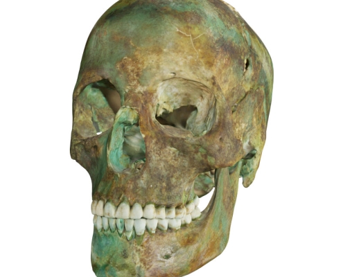
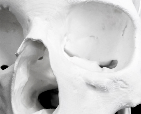
A Bronze Age skull, dating to approximately 2000 BC and belonging to the Unterwölbling culture, was used to create a manual 3D craniofacial reconstruction. The skull was unearthed during excavations at the Franzhausen cemetery I between 1981 and 1983. This location encompasses over 1200 inhumation burials, dating from 2200-2000 BC, making it a valuable resource for the study of the European Bronze Age. The skull was found in burial plot 785. Fabric remains were also discovered at the location, allowing for the reconstruction of clothing styles (Grömer, 2018). The bronze objects caused green discolouration on the bones in the burial because the calcium apatite present in bones can absorb metal elements from surrounding objects and can result in staining. Interestingly, this phenomena can help to preserve bones (Mandl, 2018).
Early Bronze Age in Lower Austria
During the Early Bronze Age (2300-1500 BC) the Únetice, Unterwölbling, and Wieselburg cultures lived in Lower Austria. The Danube river and its tributaries allowed communication between different populations and the formation of subtraditions. The most common sociopolitical organisation was based on low-level social hierarchies. Larger regional systems placed on fortified hilltops dominate the surrounding towns located near streams in valleys. The villagers had access to nonlocal metals and goods and were protected in the fortified settlements in case of war in exchange for political allegiance and obligations. Some findings suggest that rank and status are inherited patrilineally and there is a tendency for symbolic expression of power. Agriculture was the primary activity of the population, while crafts and trade ensured prosperity and hilltops were centres of trade and metal working. The Unterwölbling population lived in the southwest area of Lower Austria during the Early Bronze Age. From the valuable objects recovered in the burials at Franzhausen I, it appears that they had a key role in trading with location populations such as the Únetice and the Wieselburg cultures. The burial practices relate to the Unetice culture because they also positioned the corpes on its side facing east with north-south orientation gender differences.
Osteological Analysis
Information about the sex, ethnic group, age at death and age of bones of the skull was provided by The Natural History Museum Vienna. The skull is from a female individual aged 20-25 years that lived during the Early Bronze Age, ca. 2000 BC. An additional osteological analysis was performed for the craniofacial reconstruction by the artist to gather more information about the morphology of the skull.
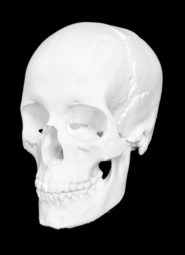
The skull has several perimortem and postmortem fractures as well as some areas missing. Most of the left and right zygomatic bones were lost and the coronoid process on the right side of the mandible is missing. Nevertheless, it was possible to create a facial reconstruction from the skull, as the trauma has caused negligible differences of the overall shape. In addition, the Department of Anthropology at the Natural History Museum Vienna indicated that during an osteological analysis that was performed on the remains for anthropological studies no plastic deformation, signs of pathologies and antemortem injuries were found on the skull.
An estimation of the sex was made following Walker’s study (2008). The smooth glabella, short mastoid process and smooth mental eminence suggested female sex. The results were further confirmed by an American/English population-specific discriminate function based on the shape of the glabella, mastoid and mental eminence. The resulting value indicated significantly that the skull is from a female individual. The results in the equation indicate male if the value is negative and female if positive.
Y = glabella X -1375 + mastoid X -1,185 + mental eminence X -1,151 + 9,128
Y = 1 X -1375 + 2 X -1,185 + 2 X -1,151 + 9,128 = 3,081
The skull is mesocephalic with average nasal spine, inferior nasal aperture with small ridge, and has a closed but visible supra nasal suture. The traverse palatine suture crosses the palate perpendicular to the median palatine suture with an anterior deviation near the juncture between the two sutures. These details suggest an individual of European ancestry (Hefner, 2009).
Methodology
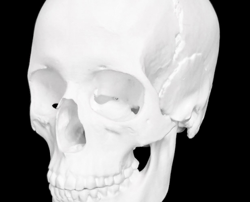
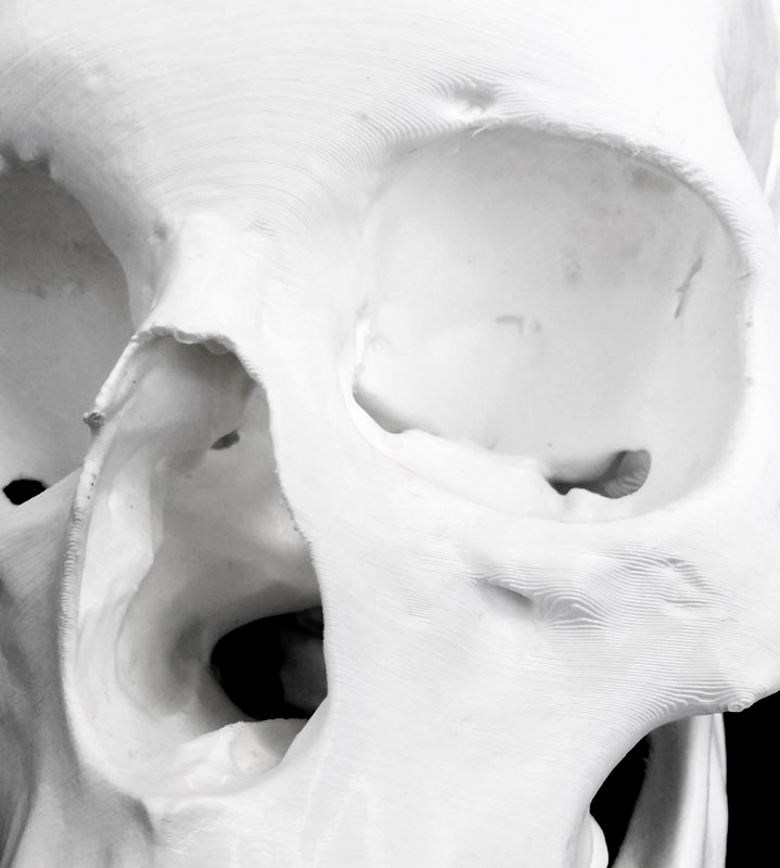
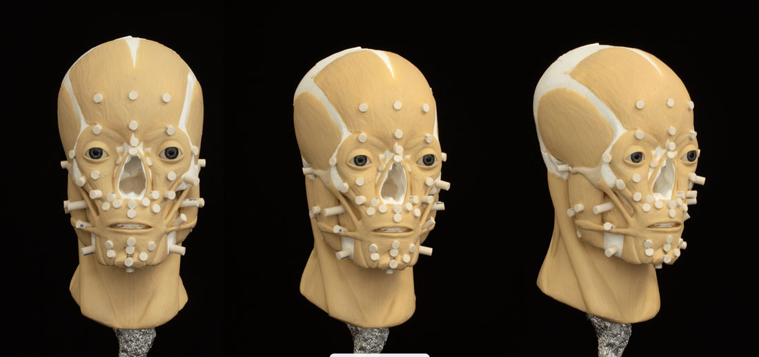
The reconstruction was made following the Manchester method. The 3D model of the skull was 3D printed and aligned with the Frankfurt Horizontal plane on a stand. The condylar processes of the mandible were placed with about 4-5mm distance from the glenoid fossa to simulate the articular disc of the temporomandibular joint (de Pontes et al, 2019). The maxillary and mandibular teeth were separated by around 2-4 mm to simulate the mandible in a rested position (Johnson et al., 2002). The mandible was also separated from the 3D model of the whole skull and was tilted slightly to be more symmetrical.
Facial soft tissue thickness (FSTT)
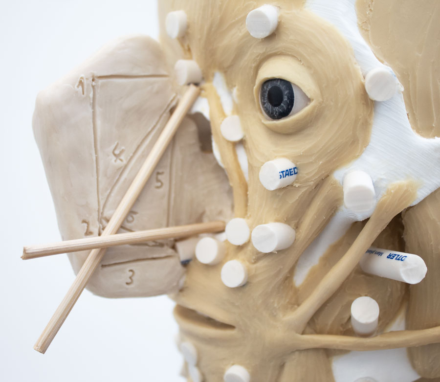
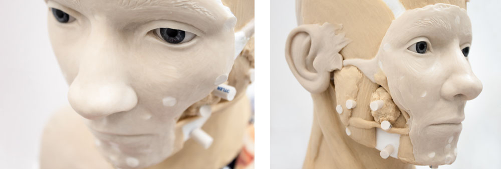
Measurements for the length of the pegs were taken from ‘Large-scale in-vivo Caucasian facial soft tissue thickness database for craniofacial reconstruction’ by De Greef et al., (2006) because this study is closest to the ancestry of the skull. The table with mean values for White adult females, aged between 18 and 29 years, with average BMI was used. The pegs were cut from cylindrical erasers and attached with glue to the skull on the landmarks outlined in the study.
Manual 3D Craniofacial Reconstruction
Eyes
Guyomarc’h et al. (2012) method determined the eye position in the frontal view and a study Wilkinson and Mautner (2003) about eye projection determined the position in the profile view.
Measurements were taken from the supraconchion to the orbitale landmarks for the orbital height and from ectoconchion to dacryon for the orbital breadth, on both sides of the skull. These measurements were then used in regression equations to determine the superiorinferior and mediolateral positions of the eyes (Guyomarc’h et al. (2012).
Right eye:
orbital height = 35 mm
orbital breadth = 37 mm
superiorinferior position = 0,54×35-3,5= 15,4mm
mediolateral position = 0,59×37-0,4= 21,43mm
Left eye:
orbital height = 36 mm
orbital breadth = 37 mm
superiorinferior position = 0,54×36-3,5= 15,94mm
mediolateral position = 0,59×37-0,4= 21,43mm
For the projection of the eyes, a tangent between the centre of supraorbital and infraorbital margins functioned as a guide. The eyes were positioned at 3.8mm past the tangent (Wilkinson and Mautner, 2003).
The position of the malar tubercle and anterior lacrimal crest determined the placement of the eye canthi. (Whitnall 1921, Balueva and Veselovskaya, 2004). The eyelid fold mirrors the shape of the supraorbital border (Balueva et al., 2009). Similarly, the eyebrow pattern mirrors the shape of the supraсiliary arch, and its inferior border closely aligns with the superior orbital ridge (Balueva et al., 2009).
A weak brow and a high nasal root suggested arched eyebrows (type ‘c’) (Fedosyutkin and Nainys, 1993; Balueva et al., 2009).
Mouth
Krogman and Iscan’s method (1986) determined the mouth width. Radiating lines from the first premolar-canine junction indicated where the mouth corners would be on the superficial skin layer. The mouth profile corresponded to the scissor-like type ‘c’ profile in Balueva and Vaselovskaya’s study (2009). The stomion was positioned according to studies by George (1993), Greyling and Meiring (1993), Ferrario et al. (2000), and Mala & Veleminska (2016).
The regression formulae from Wilkinson et al.’s (2003) research on the White European population determined the lower and upper lip heights. For these equations measurements were taken from the height of the maxillary central incisors, the height of the mandibular central incisors, and the height of the central incisors:
Upper lip height=0,4+0,6×10= 6,4mm
Lower lip height=5,5+0,4×7= 8,3mm
Tot. lip height=3,3+0,7×16,5= 14,85mm
Nose
Regression equations established by Rynn et al. (2010) and Gerasimov’s two-tangent method (1955) determined the profile shape of the nose. The angled nasal aperture and nasal tip indicated a slightly upturned nose (type ‘e’) (Rynn et al., 2010). The height of the alar groove was estimated from the position of the crista conchalis (Rynn et al., 2010).
Distances between nasion, acanthion, rhinion, and subspinale landmarks were measured for the regression equations developed by Rynn et al. (2010):
Pronasale projection perpendicular to nasion landmark (1) = 0,83×40,5-3,5= 30,12mm
Pronasale height (2) = 0,9×49-2= 42,1mm
Pronasale projection in Frankfurt Horizontal Plane (3) = 0,93×40,5-6= 31,67mm
Nasal length (4) = 0,74×51+3,5 = 41,24mm
Nasal height (5) = 0,63×51+17 = 49,13mm
Nasal depth (6) = 0,5×40,5+1,5 = 21,75mm
Studies about the relationship between the morphology of the skull and the shape of the nose by Rynn et al. (2010) and (2006) and Gerasimov (1955) determined the shape of the nose in frontal view. A regression equation for European individuals in a study by Rynn et al. (2010) was employed to estimate the maximum nasal width from the maximum nasal aperture width. The soft nasal aperture suggested the nose should be wider relative to the aperture. (Gerasimov, 1955). The marginal bifurcation of the anterior nasal spine (ANS) indicated a slightly bifurcated nasal tip (Rynn et al., 2010).
The regression equation for nasal width (Rynn et al., 2010):
MAW (maximum aperture width) = 23mm
MNW (maximum nasal width) =23×1,65 = 37,95mm
Ears
The tragus was placed above the external auditory meatus for both ears (Gatliff & Snow, 1979), with angle parallel to the angle of the posterior border of the ramus (Welcker, 1883). The nose height measurement (distance between glabella and subnasale landmarks), roughly approximates the height of the ear (Gerasimov, 1955). Due to an average 5,5mm overestimation identified in a study by Guyomarc’h and Stephan (2012), this value was subtracted from the nose height measurement.
glabella-subnasale = 63,5mm
Ear Length = 63,5-5,5= 58mm
The width of the ears was calculated from an equation in a study by Guyomarc’h and Stephan (2012).
Ear Width = 0,59 x 58 = 34,22mm
The forward angle of the mastoid indicates non-adherent lobes (Fedosyuskin and Nainys, 1993). Additionally, non-adherent lobes are common in non-Asian individuals (Guyomarc’h and Stephan, 2012).
Muscle structure
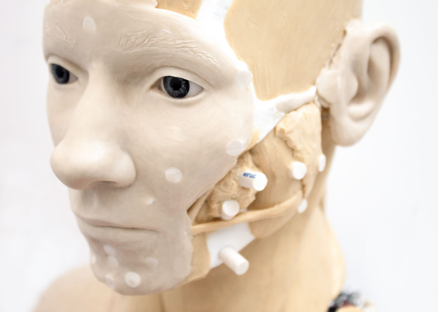
The muscles were sculpted with wax onto the skull according to the morphology of the skull and the soft tissue thickness pegs.
Skin Layer
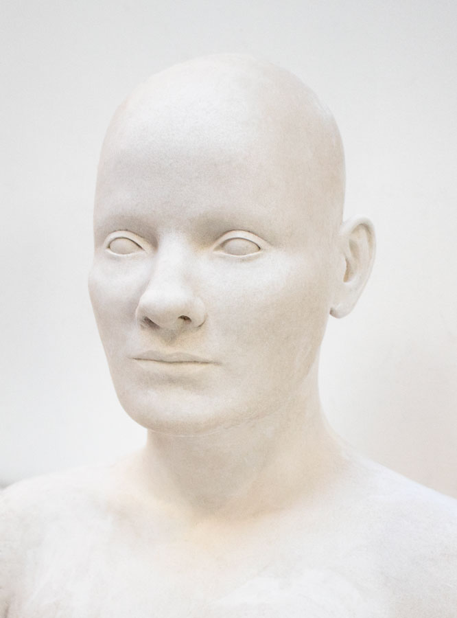
The facial features were sculpted according to the skull analysis.
Casting
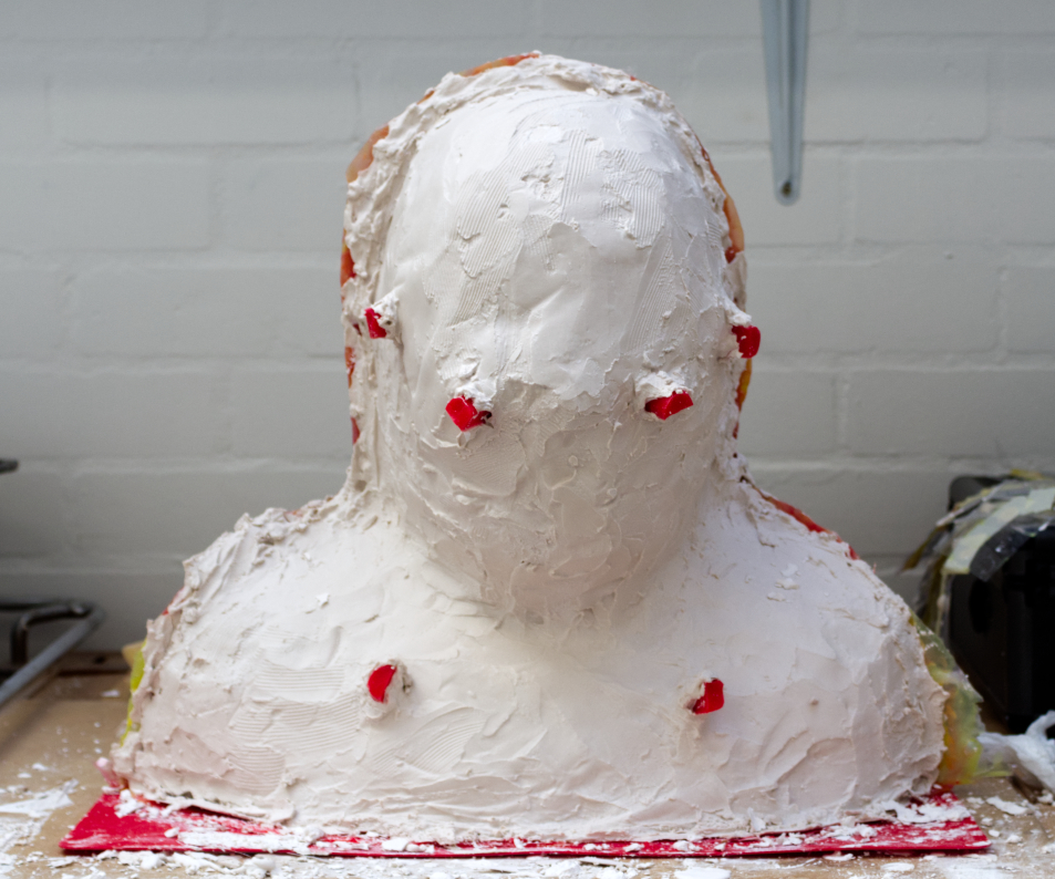
Strips of acetate sheet with keys were inserted in the wax and divided the sculpture into two haves. A 2-part mould was then made in layers of silicone and plaster, and removed from the sculpture once cured. The mould was filled with gesmonite to recreate a positive cast of the sculpture.
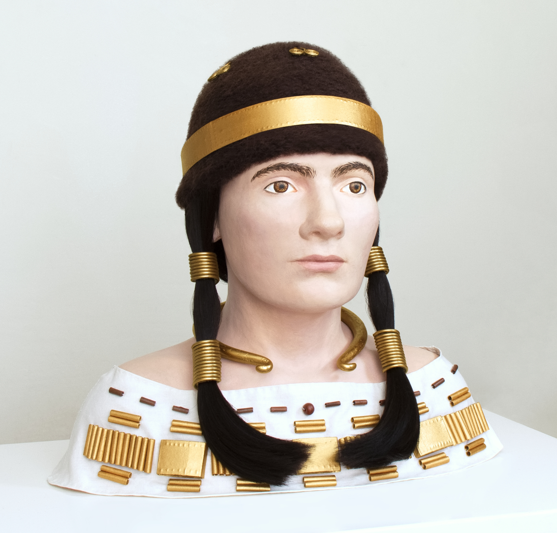
The skin texture was made with primary colours and black and white acrylics. The accessories were made of paper, cables, glue, metal wire, waxed cord and adhesive tape, then coloured with bronze paint. The upper part of the white dress was cut and sewn to fit the sculpture. The hat was made with wool. A wig was added and the accessories were sewn onto the clothes.
Final Image
References
Balueva, T., Veselovskaya, E. and Kobyliansky, E. (2009) ‘Craniofacial reconstruction by applying the ultrasound method in live human populations’, Int. J. Anthropol., vol. 24, 87–111.
De Greef at al., (2006), ‘Large-scale in-vivo Caucasian facial soft tissue thickness database for craniofacial reconstruction’, Forensic Science International
de Pontes MLC et al., (2019), ‘Correlation between temporomandibular joint morphometric measurements and gender, disk position, and condylar position’, Oral Surgery, Oral Medicine, Oral Pathology, and Oral Radiology, Vol. 128, no. 5, pp.538-542.
Fedosyutkin, B.A. and Nainys, J.V., (1993), ‘The relationship of skull morphology to facial features’, in: Iscan, M.Y. and Helmer, R.P. (eds.) Forensic analysis of the skull, New York: Wiley-Liss.
Ferrario et al., (2000), A three-dimensional quantitative analysis of lips in normal young adults, Cleft-Palate Craniofacial Journal, Vol. 37, pp. 48-54.
Gatliff, B.P. and Snow, C.C., (1979), ‘From skull to visage’, Journal of Biocommunication, Vol. 6, pp.27-30.
Greenfield, H., J., (2001), ’European Early Bronze Age’, in: Encyclopedia of Prehistory
Grömer, K., (2018), ’Visuality – Movement – Performance. The costume of a rich woman from Franzhausen in Austria, c. 2000 BC’
George, R. M., (1993), Anatomical and artistic guidelines for forensic facial reconstruction, in: Forensic Analysis of the Skull, Wiley-Liss Inc., pp. 215-27.
Gerasimov, M.M., (1955), ‘Vosstanovlenie lica po cerepu’, Moskva: Izdat. Akademii Nauk SSSR.
Greyling, I.H. and Meiring, J.H., (1993), Morphological study on the convergence of the facial muscles at the angle of the mouth, Acta Anatomica, Vol. 143, pp. 127-9.
Guyomarc’h, P. and Stephan, C.N. ,(2012), ‘The validity of ear prediction guidelines used in facial approximation’, J. Forensic Sci., vol. 57, no. 6, 1427– 1441
Guyomarc’h, P., Dutailly, B., Couture, C., and Coqueugniot, H. (2012) ‘Anatomical placement of the human eyeball in the orbit — Validation using CT scans of living adults and prediction for facial approximation’, J. Forensic Sci., vol. 57, no. 5, 1271–1275.
Hefner, J., (2009), ‘Cranial Nonmetric Variation and Estimating Ancestry’, J Forensic Sci, Vol. 54, no. 5, DOI: 10.1111/j.1556-4029.2009.01118.x
Johnson et al., (2002), ‘The determination of freeway space using two different methods, Journal of oral rehabilitation’, Vol., 29, no. 10, pp. 1010-3.
Krogman, W.M., Iscan, M.Y., (1986), ‘The Human Skeleton in Forensic Medicine’, 1st edn. Springfield, IL: C.C. Thomas Publishers.
Mala, P.Z. and Veleminska, J., (2016), ‘Vertical lip position and thickness in facial reconstruction: A validation of commonly used methods for predicting the position and size of lips’, J. Forensic Sci., vol 61, no. 4, 1046–1054.
Mandl et al., (2018), ‘The Corpse in the Early Bronze Age. Results of Histotaphonomic and Archaeothanatological Investigations of Human Remains from the Cemetery of Franzhausen I, Lower Austria’, Archaeologia Austriaca, DOI: 10.1553/archaeologia102s135.
Neave, R., (1997), ’Making Faces: Using Forensic and Archaeological Evidence’, Texas A & M University Press.
Pellegrini et al., (2011), ‘Craniofacial morphology in Austrian Early Bronze Age populations reflects sex-specific migration patterns’, Journal of Anthropological Sciences, DOI: 10.4436/jass.89013
Rynn, C., Wilkinson, C. M. and Peters, H. L., (2010), ‘Prediction of nasal morphology from the skull’, Forensic Sci. Med. Pathol., vol. 6, 20–34.
Walker, P., (2008), ’Sexing Skulls Using Discriminant Function Analysis of Visually Assessed Traits’, American Journal of physical anthropology’, Vol. 136, pp. 39-50.
Welcker, H., (1883), ‘Schiller’s Schädel und Totenmaske nebst Mittheilungen über Schädel und Totenmaske Kants’, Braunschweig: Vieweg und Sohn, pp.1-160.
Whitnall, S.E., (1921), ‘The naso-lacrimal canal: the extent to which it is formed by the maxilla, and the influence of this upon its calibre’, Ophthalmoscope, vol. 10, 557–558.
Wilkinson, C., (2004), ’Forensic Facial Reconstruction’, Cambridge University Press
Wilkinson, C.M. and Mautner, S.A., (2003), ‘Measurement of eyeball protrusion and its application in facial reconstruction’, J. Forensic Sci., vol. 48, no. 1, 12–6.
Wilkinson, C., Rynn, C., (2012), ‘Craniofacial Identification’, Cambridge University Press
Wilkinson et al., (2003), ‘The relationship between the soft tissues and the skeletal detail of the mouth’, J. Forensic Sci., vol. 48, no. 4, 728-732.


