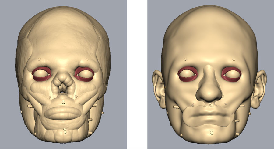
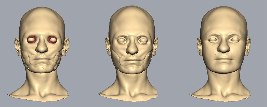
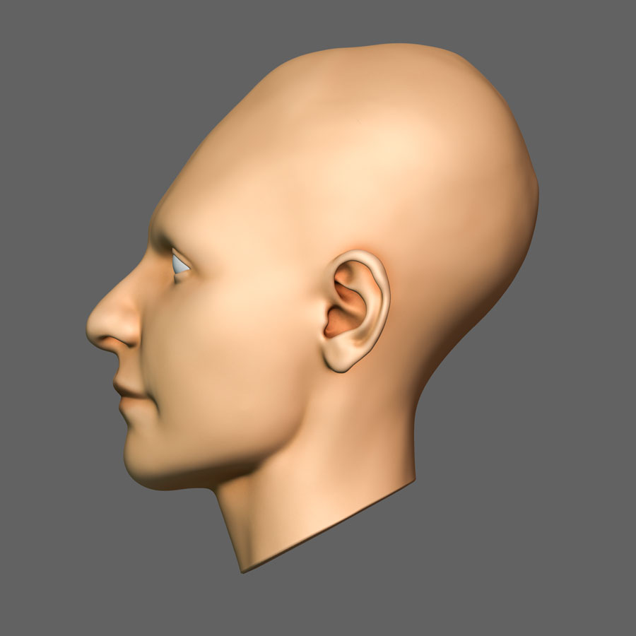
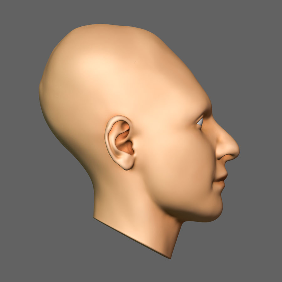
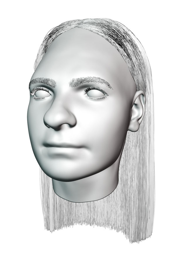
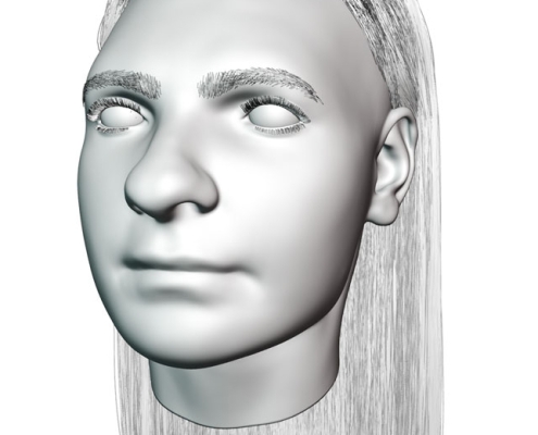
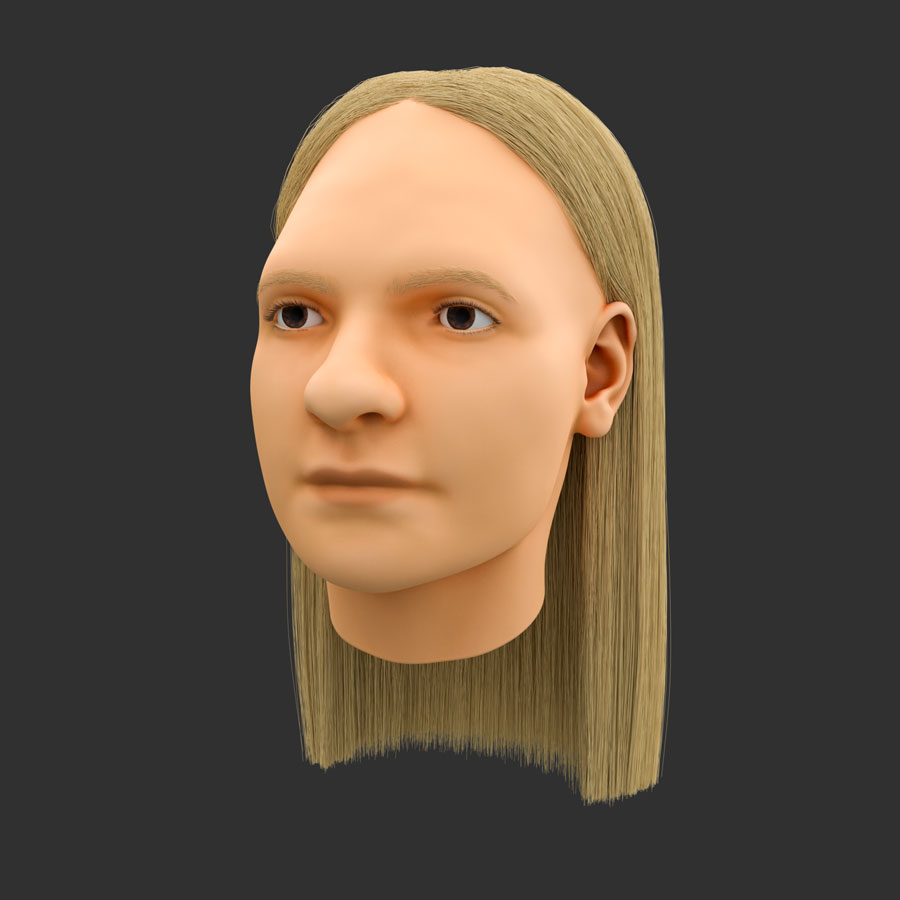
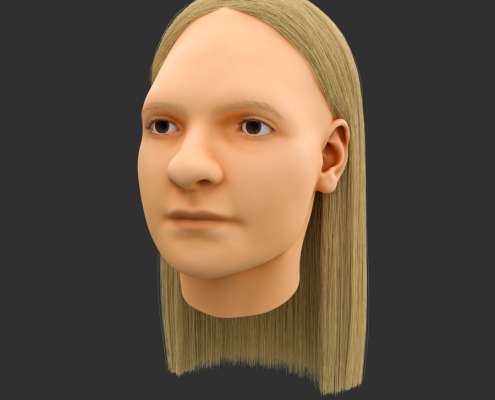
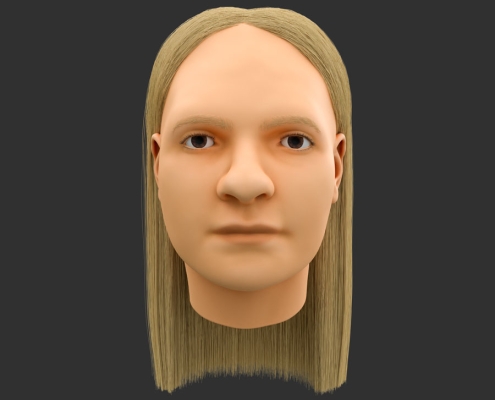
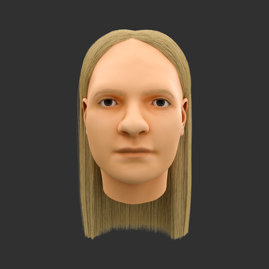
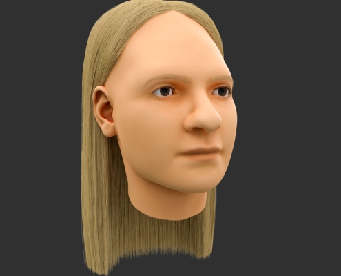
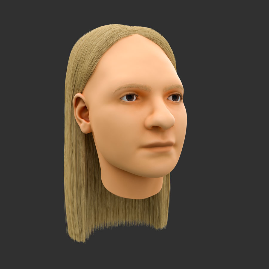
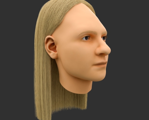
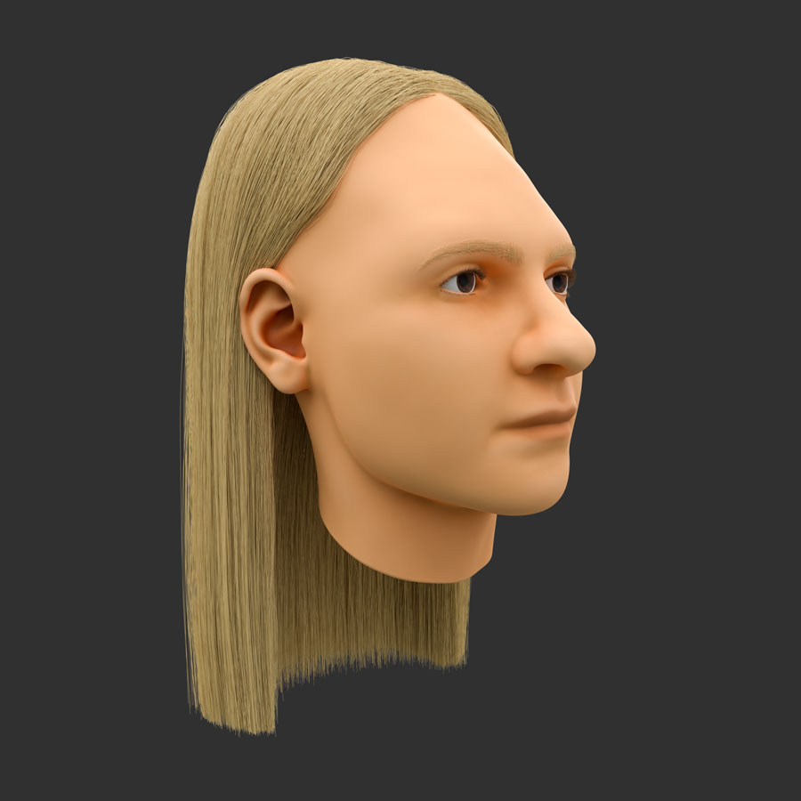
Craniofacial Reconstruction of skull from Atzgersdorf, ca. 450 CE
Cranial deformation
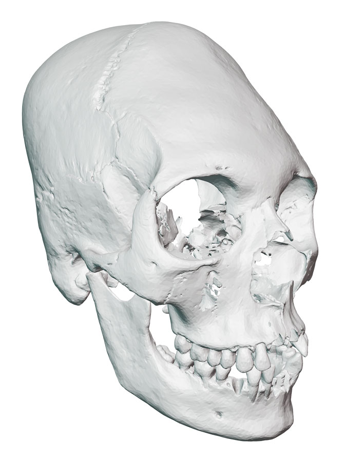
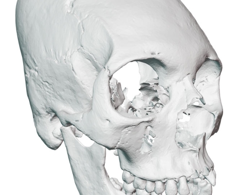
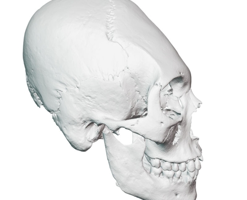
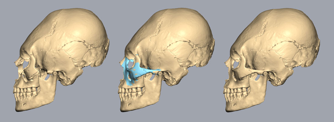
Cranial deformation, a practice of intentionally altering the shape of the skull, has a long and widespread history across the globe. Early evidence of this practice dates back to the Proto-Neolithic and Neolithic periods, with examples found in Iraq, Iran, and Syria (Meiklejohn et al., 1992). Examples from the Neolithic Age have recently been discovered also in China (Zhang, 2019). While, the Arawe population in New Britain employed this practice in recent times for aesthetic preferences (Blackwood and Danby, 1955). Cranial deformation is achieved through various methods, such as tightly wrapping the head with cloth or compressing it between flat surfaces. This compression results in an elongated skull shape. Some cultures begin this practice from birth, while others wait some months before starting. There is no anthropological evidence that suggests effects on the functioning of the brain from the deformation (Lekovic et al., 2007). This practice has been used as a way of showing status in a community, as a fashion in a culture, or as a distinguishing feature of a population. The skull used for the reconstruction shows the beauty ideals of the Huns, that incorporated cranial deformation into their culture and influenced different European ethnic groups during the Early Middle Ages. Archaeological finds from the Ostrogoths, the Lombards, the Rugii and Herulii, and the Suevi germanic tribes have been found in Austria (Schmölzer, 2016).
Osteological Analysis
Information about the sex, age at death, and age of bones of the skull was provided by The Natural History Museum Vienna. The skull is from a female individual aged 20 years that lived during the Early Medieval Age, circa 450 CE. An additional osteological analysis was performed for the craniofacial reconstruction by the artist to gather more information about the morphology of the skull.
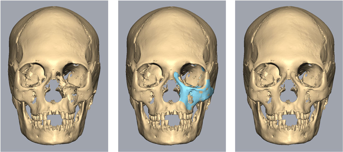
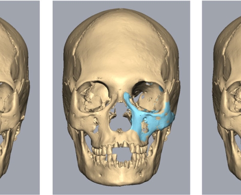
The skull has been affected by cranial deformation and is overall in a good condition, but there are some signs of damage. An anterior area on the left side of the maxillary bone is missing, several teeth such as the maxillary and mandibular central incisors, the left mandibular canine, and the right mandibular second premolar are absent, part of the left zygomatic bone is missing, the left mastoid process has been damaged, both lacrimal bones are absent and a large area of the inferior-posterior cranium has been damaged.
An estimation of the sex was made following Walker’s study (2008). The equation used is population-specific based on American/English skulls and 86,4% of the female skulls in the sample were classified correctly in the study. Skulls with scores of >0 are more likely to be from females.
Y = glabella X -1375 + mastoid X -1,185 + mental eminence X -1,151 + 9,128
Y = 2 X -1375 + 3 X -1,185 + 2 X -1,151 + 9,128 = 0,521
The result is marginally female.
Methodology
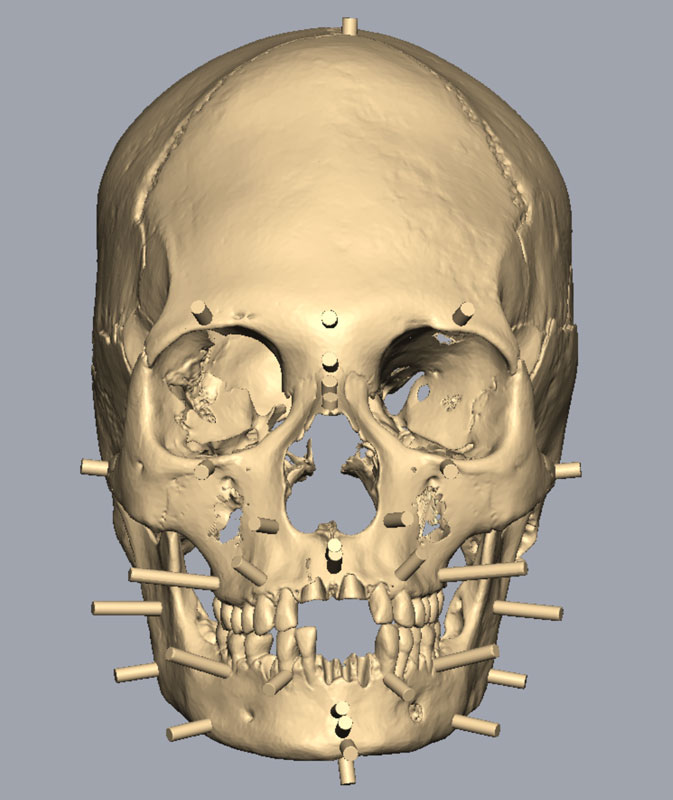
The reconstruction was made following the Manchester method. The 3D model of the skull was imported into Freeform and aligned with the Frankfurt Horizontal plane.
Skull reconstruction
The anterior left side of the maxillary bone was reconstructed. The complete right side was isolated and mirrored in Blender, then imported into Freeform where it was further adjusted with the tug tool and merged with the skull.
Facial soft tissue thickness (FSTT)
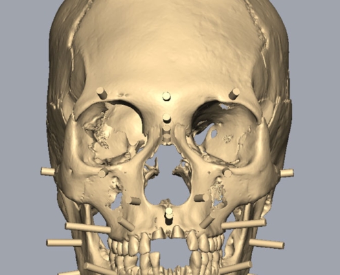
Measurements for the length of the pegs were taken from the 2018 tallied facial soft tissue thicknesses table devised for adults, which was established by Stephan (2017). The table in this study integrates all previously published FSTT literature to establish a non-population specific dataset that comprises a large sample size. This study was used because the ancestry of the skull was less specific compared to the skull from Franzhausen. In addition, the greater sample size used in Stephan’s (2017) study presents less sample bias, with any errors in measurement mode and method being averaged out.
Eyes
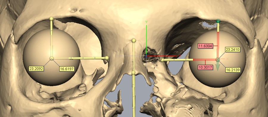
Guyomarc’h et al. (2012) method determined the position of the eyes in frontal view and a study by Wilkinson and Mautner (2003) about eye projection determined the position in profile view.
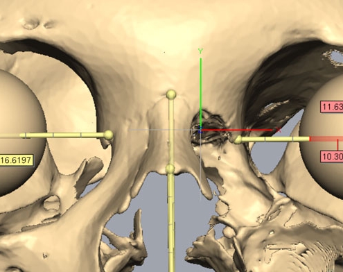
Measurements were taken of distance between the supraconchion and the orbitale landmarks for the orbital height and of distance between ectoconchion and dacryon for the orbital breadth on both sides of the skull. These measurements were then used in regression equations to determine the superiorinferior and mediolateral positions of the eyes (Guyomarc’h et al. (2012).
Right eye:
orbital height = 37,25 mm
orbital breadth = 40 mm
superiorinferior position = 0,54×37,25-3,5= 16,62mm
mediolateral position = 0,59×40-0,4= 23,2mm
Left eye:
orbital height = 36,7 mm
orbital breadth = 40 mm
superiorinferior position = 0,54×36,7-3,5= 16,32mm
mediolateral position = 0,59×40-0,4= 23,2mm
For the projection of the eyes a tangent between the centre of supraorbital and infraorbital margins functioned as a guide. The eyes were positioned at 3.8mm past the tangent (Wilkinson and Mautner, 2003).
The positioned of the malar tubercle and anterior lacrimal crest determined the placement of the eye canthi. (Whitnall 1921, Balueva and Veselovskaya, 2004). The eyelid fold mirrors the shape of the supraorbital border (Balueva et al., 2009). Similarly, the eyebrow pattern mirrors the shape of the supraciliary arch and its lower edge is placed on top of the orbital border (Balueva et al., 2009).
A weak brow and a high nasal root, suggested arched eyebrows (type ‘c’). (Fedosyutkin and Nainys, 1993; Balueva et al., 2009).
Mouth
Krogman and Iscan’s method (1986) determined the mouth width. Radiating lines from the first premolar-canine junction indicated where the mouth corners would be on the superficial skin layer. The mouth profile corresponded to the scissor-like ‘type c’ profile in Balueva and Vaselovskaya’s study (2009). The stomion was positioned according to studies by George (1993), Greyling and Meiring (1993), Ferrario et al. (2000), and Mala & Veleminska (2016). It was not possible to use the regression formulas by Wilkinson et al. (2003) because both mandibular and maxillary central incisors were missing.
Nose
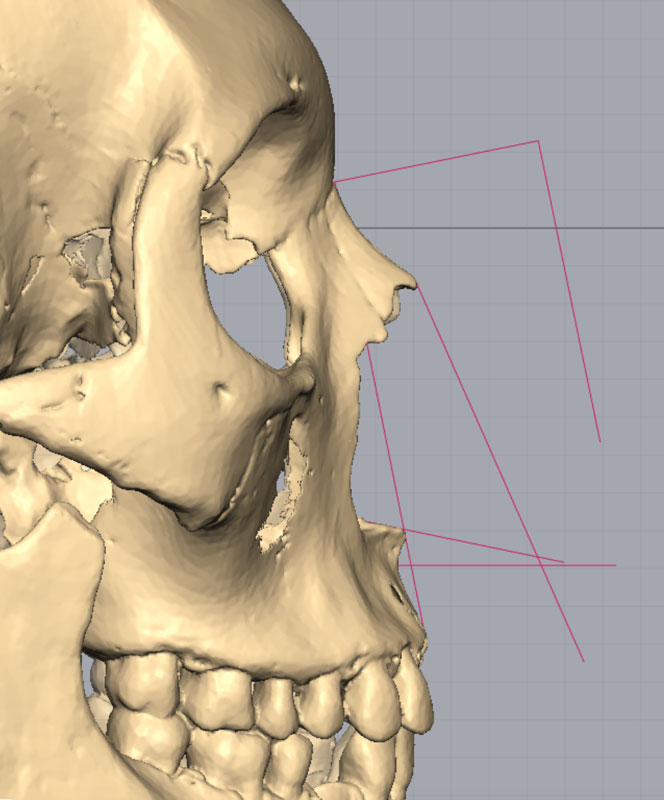
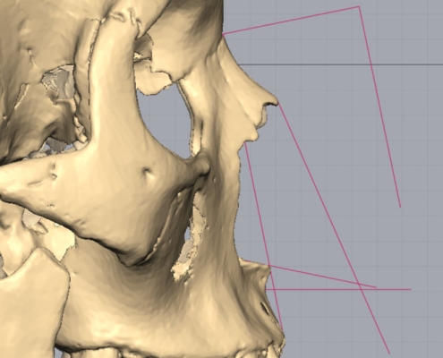
Regression equations developed by Rynn et al. (2010) and Gerasimov’s two-tangent method (1955) determined the profile shape of the nose. The angle of the nasal spine indicated a slightly upturned nose (type ‘e’) (Rynn et al., 2010). The height of the alar groove was estimated from the position of the crista conchalis (Rynn et al., 2010).
Distances between nasion, acanthion, rhinion, and subspinale landmarks were measured for the regression equations developed by Rynn et al. (2010):
Pronasale projection perpendicular to nasion landmark (1) = 0,83×37,53-3,5= 27,65mm
Pronasale height (2) = 0,9×47,47-2= 40,72mm
Pronasale projection in Frankfurt Horizontal Plane (3) = 0,93×37,53-6= 28,9mm
Nasal length (4) = 0,74×51,61+3,5 = 41,69mm
Nasal height (5) = 0,63×51,61+17 = 49,51mm
Nasal depth (6) = 0,5×37,53+1,5 = 20,27mm
Studies on the relationship between the morphology of the skull and the shape of the nose by Rynn et al. (2006, 2010) and Gerasimov (1955) determined the shape of the nose in frontal view. A regression equation for European individuals in a study by Rynn et al. (2010) was employed to estimate the maximum nasal width from the maximum nasal aperture width. The soft nasal aperture suggested the nose should be wider relative to the aperture. (Gerasimov, 1955). The bifurcated shape of the anterior nasal spine (ANS) indicated a bifurcated nasal tip (Rynn et al., 2010).
Regression equation for nasal width (Rynn et al., 2010):
MAW (maximum aperture width) = 25,4mm
MNW (maximum nasal width) =25,4×1,65 = 41,91mm
Ears
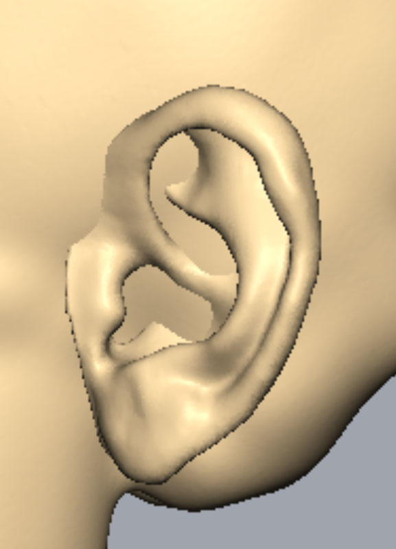
The ears were positioned on the skull with the external auditory meatus at the top of the tragus (Gatliff & Snow, 1979), with angle parallel to the angle of the posterior border of the ramus (Welcker, 1883). The nose height measurement (distance between glabella and subnasale landmarks), roughly approximates the height of the ear (Gerasimov, 1955) with a 5,5mm overestimation identified in a study by Guyomarc’h and Stephan (2012) that was subtracted from the measurement.
glabella-subnasale = 63,78mm
Ear Length = 63,78-5,5= 58,28mm
The width of the ears was calculated from an equation in a study by Guyomarc’h and Stephan (2012).
Ear Width = 0,59 x 58,28 = 34,39mm
The forward angle of the mastoid indicates non-adherent lobes (Fedosyuskin and Nainys, 1993). Additionally, non-adherent lobes are common in non-Asian individuals (Guyomarc’h and Stephan, 2012).
Muscle structure
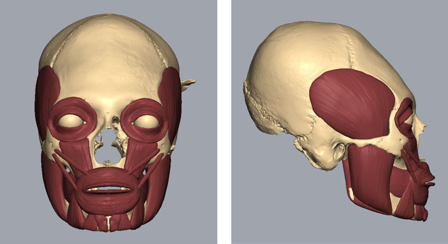
The muscle structure was positioned and shaped according to the morphology of the skull.
Skin layer
All the muscles (except for the orbicularis oculi) and the skull where merged together and a 6.5 mm offset layer was created in Freeform to mimic the thickness of the skin. The new layer was then smoothed. Facial features of scans from individuals were positioned and shaped according to the skull analysis. Then the subcutaneous fat was added and smoothed and the neck and eyebrows were added. The layers of the soft tissue were merged together and exported into an obj 3D model, then imported into Blender where further adjustments were made.
Texture and hair
The skin texture was created in blender with procedural nodes in the shading editor and manual painting in photoshop. The texture for the eyes was made in photoshop.
Hair, eyebrows and eyelashes were added to the model with the new hair system based on curves in blender version 3.3 and coloured in the shading editor.
Final Images
References
Balueva, T., Veselovskaya, E. and Kobyliansky, E. (2009) ‘Craniofacial reconstruction by applying the ultrasound method in live human populations’, Int. J. Anthropol., vol. 24, 87–111.
Blackwood, B., Danby, P., M., (1955), ‘A Study of Artificial Cranial Deformation in New Britain’, The Journal of the Royal Anthropological Institute of Great Britain and Ireland, Vol. 85, No. 1/2, pp. 173-191.
de Pontes MLC et al., (2019), ‘Correlation between temporomandibular joint morphometric measurements and gender, disk position, and condylar position’, Oral Surgery, Oral Medicine, Oral Pathology, and Oral Radiology, Vol. 128, no. 5, pp.538-542.
Fedosyutkin, B.A. and Nainys, J.V., (1993), ‘The relationship of skull morphology to facial features’, in: Iscan, M.Y. and Helmer, R.P. (eds.) Forensic analysis of the skull, New York: Wiley-Liss.
Ferrario et al., (2000), A three-dimensional quantitative analysis of lips in normal young adults, Cleft-Palate Craniofacial Journal, Vol. 37, pp. 48-54.
Gatliff, B.P. and Snow, C.C., (1979), ‘From skull to visage’, Journal of Biocommunication, Vol. 6, pp.27-30.
George, R. M., (1993), Anatomical and artistic guidelines for forensic facial reconstruction, in: Forensic Analysis of the Skull, Wiley-Liss Inc., pp. 215-27.
Gerasimov, M.M., (1955), ‘Vosstanovlenie lica po cerepu’, Moskva: Izdat. Akademii Nauk SSSR.
Greyling, I.H. and Meiring, J.H., (1993), Morphological study on the convergence of the facial muscles at the angle of the mouth, Acta Anatomica, Vol. 143, pp. 127-9.
Guyomarc’h, P. and Stephan, C.N. ,(2012), ‘The validity of ear prediction guidelines used in facial approximation’, J. Forensic Sci., vol. 57, no. 6, 1427– 1441.
Guyomarc’h, P., Dutailly, B., Couture, C., and Coqueugniot, H. (2012) ‘Anatomical placement of the human eyeball in the orbit — Validation using CT scans of living adults and prediction for facial approximation’, J. Forensic Sci., vol. 57, no. 5, 1271–1275.
Hefner, J., (2009), ‘Cranial Nonmetric Variation and Estimating Ancestry’, J Forensic Sci, Vol. 54, no. 5, DOI: 10.1111/j.1556-4029.2009.01118.x
Johnson et al., (2002), ‘The determination of freeway space using two different methods, Journal of oral rehabilitation’, Vol., 29, no. 10, pp. 1010-3.
Krogman, W.M., Iscan, M.Y., (1986), ‘The Human Skeleton in Forensic Medicine’, 1st edn. Springfield, IL: C.C. Thomas Publishers.
Lekovic et al., (2007), ’New World cranial deformation practices: historical implications for pathophysiology of cognitive impairment in deformational plagiocephaly’, Neurosurgery, Vol. 60, No. 6, pp1137-46, DOI: 10.1227/01.NEU.0000255462.99516.B0
Mala, P.Z. and Veleminska, J., (2016), ‘Vertical lip position and thickness in facial reconstruction: A validation of commonly used methods for predicting the position and size of lips’, J. Forensic Sci., vol 61, no. 4, 1046–1054.
Meiklejohn et al., (1992), ’Artificial cranial deformation in the Proto-Neolithic and Neolithic Near East and its possible origin: Evidence from four sites’, Paléorient, Vol. 18, No. 2, pp. 83-97, DOI: 10.3406/paleo.1992.4574
Neave, R., (1997), ’Making Faces: Using Forensic and Archaeological Evidence’, Texas A & M University Press.
Rynn, C., Wilkinson, C. M. and Peters, H. L., (2010), ‘Prediction of nasal morphology from the skull’, Forensic Sci. Med. Pathol., vol. 6, 20–34.
Schmölzer, A., (2016), ‘The Long-Heads – Strangers of the East? Artificial Cranial Deformation in Austria’, in: ‘Through the Eyes of a Stranger: Appropriating the Foreign and Transforming the Local Context’.
Stephan, C., (2017), ’2018 tallied facial soft tissue thicknesses for adults and sub-adults’, Forensic Science International, Vol. 280, 113-123.
Walker, P., (2008), ’Sexing Skulls Using Discriminant Function Analysis of Visually Assessed Traits’, American Journal of physical anthropology’, Vol. 136, pp. 39-50.
Welcker, H., (1883), ‘Schiller’s Schädel und Totenmaske nebst Mittheilungen über Schädel und Totenmaske Kants’, Braunschweig: Vieweg und Sohn, pp.1-160.
Whitnall, S.E., (1921), ‘The naso-lacrimal canal: the extent to which it is formed by the maxilla, and the influence of this upon its calibre’, Ophthalmoscope, vol. 10, 557–558.
Wilkinson, C., (2004), ’Forensic Facial Reconstruction’, Cambridge University Press.
Wilkinson, C.M. and Mautner, S.A., (2003), ‘Measurement of eyeball protrusion and its application in facial reconstruction’, J. Forensic Sci., vol. 48, no. 1, 12–6.
Wilkinson, C., Rynn, C., (2012), ‘Craniofacial Identification’, Cambridge University Press.
Wilkinson et al., (2003), ‘The relationship between the soft tissues and the skeletal detail of the mouth’, J. Forensic Sci., vol. 48, no. 4, 728-732.
Zhang et al., (2019), ’Intentional cranial modification from the Houtaomuga Site in Jilin, China: Earliest evidence and longest in situ practice during the Neolithic Age’, Am J Phys Anthropol, 169: 747–756, DOI: 10.1002/ajpa.23888

